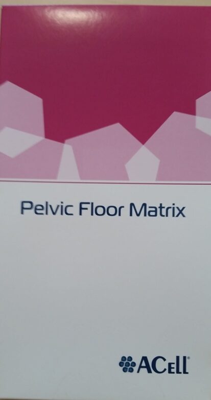
Matristem Pelvic Floor Matrix

Miscellaneous Pfm0912 Acell Matristem Pelvic Floor Matrix 9cm X 12cm Esutures
Acell Pfm0710 Pelvic Floor Matrix 7 X 10cm Medical Equipment Export

Acell Initiates Post Market Study Of Matristem Pelvic Floor Matrix

Matristem Product List Call 1 800 826 2926 Or Acell 60mg Matristem Micromatrix Micronized Particles 5 Box Mm0100 100mg Matristem Particles Fine 1 Box Mm0200 200mg Matristem

Matrix Lar Pelvic Acell Pfm0710 7 X 10cm As Mhalairt Uidheamachd Meidigeach

Towards Rebuilding Vaginal Support Utilizing An Extracellular Matrix Bioscaffold Sciencedirect
The primary objective of this study is to assess the safety and effectiveness of matristem pelvic floor matrix as compared to native tissue repair for the treatment of pelvic organ prolapse.
Matristem pelvic floor matrix.

Wound Matrix Products Medline Industries Inc

Figure 2 From Extracellular Matrix Regenerative Graft Attenuates The Negative Impact Of Polypropylene Prolapse Mesh On Vagina In Rhesus Macaque Semantic Scholar

Cytal Burn Matrix Acell
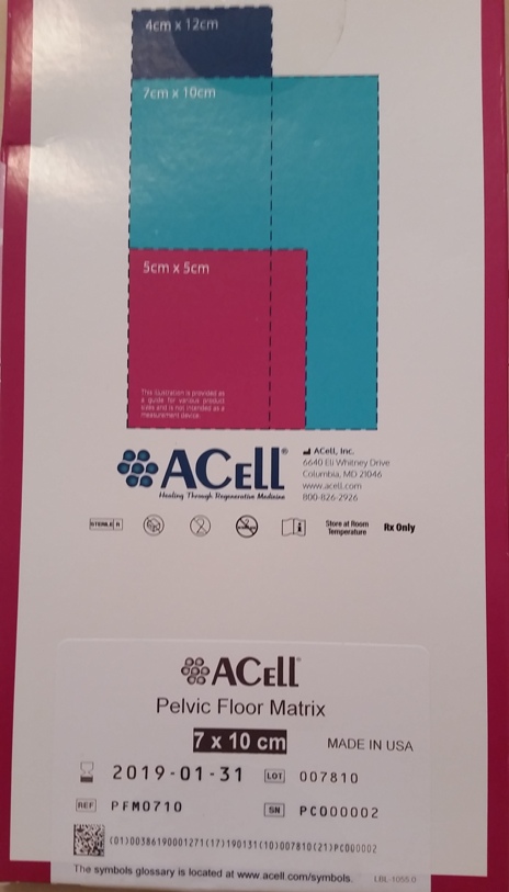
Acell Pfm0710 Pelva Planta Matrico 7 X 10cm Eksportado Pri Kuracaj Ekipaĵoj

Impact Of Polypropylene Prolapse Mesh On Vaginal Smooth Muscle In Rhesus Macaque Sciencedirect
Pdf Recent Advances In Pelvic Floor Repair

Extracellular Matrix As A Bioscaffold For Tissue Engineering Sciencedirect

Heather Van Raalte Md Princeton Urogyn
Https Www Fda Gov Media 122854 Download

Pdf Biomaterials For Pelvic Floor Reconstructive Surgery How Can We Do Better
Acell Matristem Presentation By Michael Snyder

Extracellular Matrix Regenerative Graft Attenuates The Negative Impact Of Polypropylene Prolapse Mesh On Vagina In Rhesus Macaque American Journal Of Obstetrics Gynecology

Remodeling Response An Overview Sciencedirect Topics
Https Www Ics Org Workshops Handoutfiles 000749 Pdf

The Role Of Skin Substitutes In The Management Of Chronic Cutaneous Wounds Greaves 2013 Wound Repair And Regeneration Wiley Online Library
Https Www Fepblue Org Media Pdfs Medical 20policies 04 10 2020 701113 20bioengineered 20skin 20and 20soft 20tissue 20substitutes Pdf
Https Www Aapc Com Localchaptereventagendas Ba1e2f30 94dc 49a2 9b23 D5230d1ab3cc Pdf
Https Media Fepblue Org Media F3d086ed611841ea814af0c07c87866d Pdf

Presence Of Relaxin 2 Oxytocin And Their Receptors In Uterosacral Ligaments Of Pre Menopausal Patients With And Without Pelvic Organ Prolapse Request Pdf
Source : pinterest.com
