May Appear On A Radiograph As An Elevated Bladder Floor

Pin By Diana Diana On Pelvic Enlarged Prostate Renal Stones Bladder

Scoliosis Jpg 680 539 Pixels Massage Therapy Scoliosis Scoliosis Exercises

Assessment Of Pelvic Organ Prolapse A Review Shek 2016 Ultrasound In Obstetrics Amp Gynecology Wiley Online Library

Ultrasound In The Investigation Of Posterior Compartment Vaginal Prolapse And Obstructed Defecation Dietz 2012 Ultrasound In Obstetrics Amp Gynecology Wiley Online Library

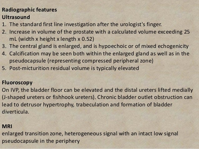
Ultrasound Imaging In Assessment Of The Male Patient With Voiding Dysfunction Radiology Key
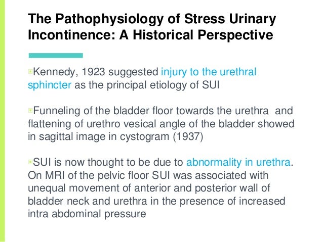
Stress Urinary Incontinence

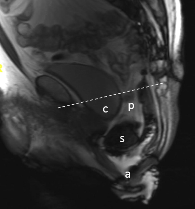
Recurrent prolapse in a patient who had previously undergone hysterectomy and more recently urinary bladder suspension for urinary incontinence.
May appear on a radiograph as an elevated bladder floor.

Pelvic Floor Laxity A Not So Rare But Unrecognized Form Of Daytime Urinary Incontinence In Peripubertal And Adolescent Girls Journal Of Pediatric Urology

Pathogenesis Of Urethral Funneling In Women With Stress Urinary Incontinence Assessed By Introital Ultrasound Tunn 2005 Ultrasound In Obstetrics Amp Gynecology Wiley Online Library

Bladder And Ureteral Imaging Radiology Key

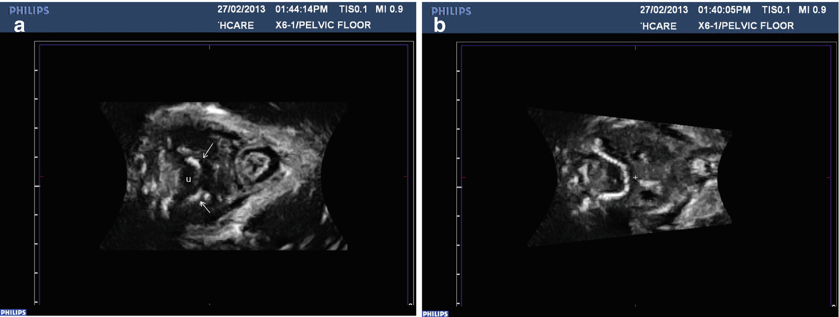
Pdf Assessment Of Pelvic Floor Movement Using Transabdominal And Transperineal Ultrasound
Https Scholarcommons Usf Edu Cgi Viewcontent Cgi Article 6484 Context Etd

Pin On Nursing

Renal Calculi Kidney Stones Concepts For Registered Nurse Not That I Don T Know These Being On A Ur Nursing School Survival Renal Calculi Nursing Mnemonics
Imaging Of Chronic Male Pelvic Pain What The Abdominal Imager Should Know Springerlink

Pin On Pudendal Nerve

Pdf Ultrasound Of The Urinary Bladder Revisited

Pelvic Brim Demarcation Of True And False Pelvis Sacroiliac Joint Sacroiliac Pelvis

Pin On Pelvic Floor
Pelvic Floor Ultrasound Springerlink

References In Magnetic Resonance Imaging Of The Female Pelvic Floor Radiologic Clinics
Gut

Pelvic Floor Exercises Virginia Urology

Necrotizing Fasciitis Bing Images Body Disorders Dermatology Informative
Https Onlinelibrary Wiley Com Doi Pdf 10 1002 Nau 23426

Uterus Fascial Ligaments Anatomy Round Ligament Of Uterus Uterine Fallopian Tube Proper Ovarian Ligament Ligament Of Ova In 2020 Uterus Fallopian Tubes Anatomy
Source : pinterest.com
